New microscope shows live cells in 3 dimensions
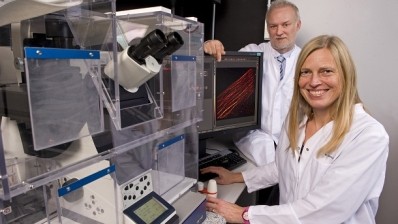
Nanolive, got its start at the EPFL Innovation Park in Lausanne in 2013 and has since developed the 3D Cell Explorer microscope, which can reveal living cells without dyes, chemical reagents or genetic modifications.
The microscope will be on the market this summer, and the company has already received over 50 preorders according to an item on news-medical.net. Early orders are apparently discounted, but the machine will soon sell for €19,900 a pop.
Tech talk
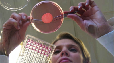
Tomography is the sort of imaging technology that facilitates MRIs and CT scans. Besides being able to show live 3D images, this method of inspection means that the cells and their functionality can be seen as is: “As no chemistry or marker is used at all, the observation is completely non-invasive to the cell, and allows resolving the cell’s parts down to 200nm,” reports news-medical.net.
The resulting images, shown on a computer screen, are “complete tomography of the refractive index within the living cell.”
Researchers using the new technology, “will actually see and measure precisely the impact of stimuli and drugs on cells, thus enabling completely new fields of research and smarter products,” says Nanolive.
Software capabilities
For new technologies to integrate effectively in the marketplace, they need to not only move things forward in dynamic ways but also be in ready conversation with established methods. This eases adoption and allows researchers to continue with their current projects while discovering what else is now possible.
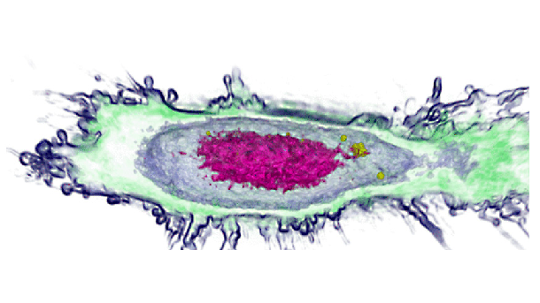
That seems to be the logic behind the software accompanying Nanolive’s 3D Cell Explorer. “To mark and label certain parts of the measured cells in 3D, the software allows the user to ‘virtually brush’ and ‘digitally paint’ the part of a cell based on their physical properties,” explains news-medical.net.
The software, known as STEVE, tracks the digital staining as it happens in 3D as well as in 2D. Researchers can adjust the coloring and reuse a palette for related cell images.
Research potential
Faith Troy, scientific advisor, and Yann Cotte, CEO, founded the company, and Nanolive has since become a team of 11.
The company calls the 3D Cell Explorer “a major milestone in the history of microscopy, which may change all the rules in the fields of Education, Biology, Pharmaceutics, Cosmetics, Labs and Industry.”
Applications of the technology include, cell morphology monitoring, differentiation of over 200 cell types, cell-cell interaction, intracellular trafficking, cellular remodeling processes, apoptosis, autophagy, necrosis, cell proliferation, nanoparticle internalization, and more, says Nanolive.